Learning Outcomes:
i. Describe the key components of skeletal muscle at the cellular level, including muscle fibers, sarcomeres, and myofibrils.
ii. Explain the role of actin, myosin, and other proteins in muscle contraction.
iii. Understand how the intricate arrangement of these structures allows skeletal muscle to generate force and movement.
Introduction:
Imagine a mighty athlete preparing for a race. Their muscles, unseen to the naked eye, are a bustling city of microscopic machines ready to generate explosive power. In this lesson, we'll shrink down to explore the ultra-structure of skeletal muscle, unveiling the hidden world that drives every movement we make.
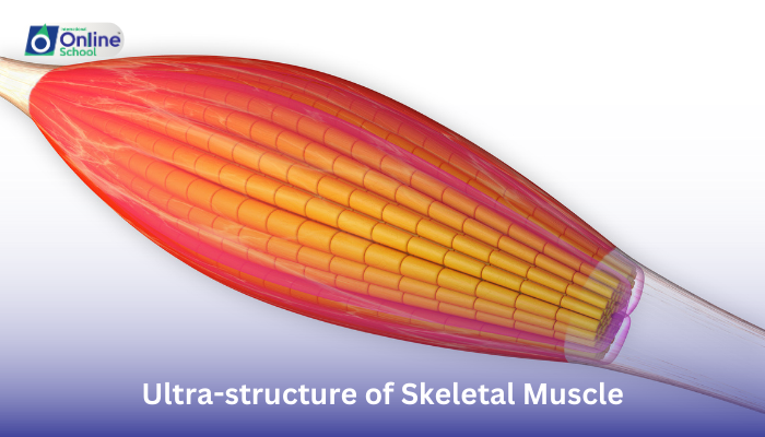
i. Muscle Fibers: The Powerhouse Units:
Our journey begins with muscle fibers, the long, cylindrical cells that make up skeletal muscle. Each fiber is a self-contained powerhouse, packed with specialized structures called myofibrils, the true engines of movement.
ii. Sarcomeres: The Contractile Champs:
Myofibrils are further organized into repeating units called sarcomeres, which look like striped ribbons under a microscope. These sarcomeres are the fundamental units of muscle contraction, containing two main types of protein filaments:
Actin: These thin filaments, like tiny ropes, run parallel to each other within the sarcomere.
Myosin: These thicker filaments, like molecular motors, are arranged between the actin filaments.
iii. The Dance of Actin and Myosin:
When a muscle receives a signal from the nervous system, a fascinating dance begins:
Calcium triggers the action: Calcium ions flood into the sarcomere, activating the myosin motors.
Myosin reaches for actin: The "heads" of the myosin molecules attach themselves to the actin filaments.
Myosin pulls, actin slides: Powered by its internal engine, each myosin head pulls the actin filament towards itself, shortening the sarcomere.
Force generation and movement: As all the myosin heads pull together, the entire myofibril shortens, generating the force that ultimately contracts the muscle fiber and allows for movement.
iv. The Supporting Cast:
This remarkable performance of actin and myosin is supported by numerous other proteins:
Tropomyosin: This regulatory protein covers the actin filaments, preventing myosin from binding unless activated by calcium.
Troponin: This protein complex works with calcium to control the interaction between actin and myosin, regulating the timing and strength of muscle contraction.
Sarcoplasmic reticulum: This specialized membrane network stores and releases calcium ions, playing a crucial role in triggering and regulating muscle contraction.
v. A Symphony of Teamwork:
The intricate arrangement and coordinated action of these proteins within the sarcomere is what allows skeletal muscles to generate the force and movement we need for everything from lifting a feather to running a marathon.
Exploring the ultra-structure of skeletal muscle reveals a hidden world of microscopic choreography, where proteins like actin and myosin orchestrate the powerful dance of contraction. By understanding these intricate details, we gain a deeper appreciation for the remarkable engineering and functionality of our muscles, the unseen heroes that fuel every movement and allow us to experience the world in all its dynamic glory. So, the next time you flex your arm or take a step, remember the silent symphony of muscle fibers and sarcomeres that powers every action, a testament to the wonders that lie within the human body.
